

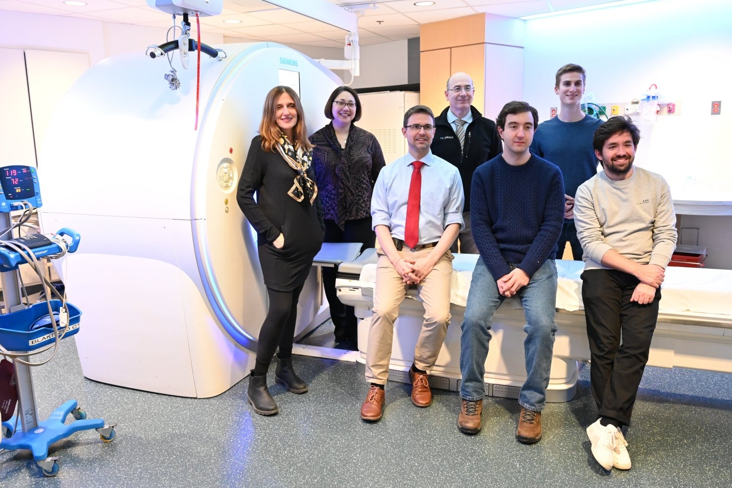
The name Sybil has its origins in the oracles of ancient Greece, also known as Sibyls: female figures relied upon to impart divine knowledge of the unseen, the past, the Almighty, the present, and the future. Now, the name has been mined from ancient times and given to an artificial intelligence tool for lung cancer risk assessment being developed by researchers at MIT’s Abdul Latif Jameel Machine Learning Clinic in Health, General Comprehensive Cancer Center (MGCC), and Chang Gung Memorial Hospital. (CGMH).
Lung cancer is the #1 deadliest cancer in the world, killing 1.7 million people worldwide in 2020, killing more people than the next three deadliest cancers combined.
“It is the biggest killer of cancer because it is relatively common and relatively difficult to treat, especially once it has reached an advanced stage,” says Florian Ventelmann, MD, a thoracic interventional radiologist at MGCC and a co-author on the new work. “In this case, it is important to know that if you detect lung cancer early, the long-term outcome is much better. The five-year survival rate is close to 70 percent, whereas if you detect it when it is advanced, the two-year survival rate is Five years it’s just under 10 percent.”
Although there has been an increase in new therapies introduced to combat lung cancer in recent years, the majority of lung cancer patients still undergo this disease. Low-dose computed tomography (LDCT) scans of the lung are currently the most common way to screen patients for lung cancer in hopes of finding it in the early stages, when it can still be surgically removed. Sybil takes screening a step further, analyzing LDCT image data without the help of a radiologist to predict a patient’s future risk of developing lung cancer within six years.
In their new paper published in Journal of Clinical OncologyResearchers at Jameel Clinic, MGCC, and CGMH showed that Sybil obtained C-indices of 0.75, 0.81, and 0.80 over six years from various sets of lung LDCT scans taken from the National Lung Cancer Screening Trial (NLST), a comprehensive public trial. Hospital (MGH) and CGMH, respectively – Models achieving a C-index score above 0.7 are considered good and over 0.8 are considered strong. The ROC-AUCs for 1-year prediction using Sybil scored higher, ranging from 0.86 to 0.94, with 1.00 being the highest possible score.
Although successful, the 3D nature of a lung CT scan made Sybil a challenge to build. Co-author Peter Michael, an MIT PhD student in electrical engineering and computer science affiliated with the Jameel Clinic and MIT Computer Science and Artificial Intelligence Laboratory (CSAIL), likened the process to “trying to find a needle in a haystack.” The imaging data used to train Sybil was largely absent of any signs of cancer because early-stage lung cancer occupies small portions of the lung—just a fraction of the hundreds of thousands of pixels that make up each CT scan. The denser portions of lung tissue are known as pulmonary nodules, and although they may be cancerous, most are not, and can occur from healed infections or airborne irritants.
To ensure that Sybil will be able to accurately assess cancer risk, Fintelmann and his team have labeled hundreds of CT scans with visible cancerous tumors that can be used to train Sybil before testing the model on a CT scan without visible signs of cancer.
MIT electrical engineering and computer science doctoral student Jeremy Wolfend, co-author of the paper and Jameel Clinic and subsidiary of CSAIL, was surprised by how high Sybil’s scores were despite not having any visible cancer. “We found that while we[as humans]can’t quite pinpoint where the cancer is, the model can still have some predictive power about which lung will eventually develop cancer,” he recalls. “Knowing (Sybil) was able to highlight the most likely aspect of it was really interesting for us.”
Co-author Lecia V. Sequist, a medical oncologist, lung cancer expert and director of the Center for Innovation in Early Cancer Detection at MGH, says the results achieved by the team with Sybil are significant “because lung cancer screening has not yet been rolled out to its fullest potential in the United States or globally.” world, and Sybil may be able to help us bridge that gap.”
Lung cancer screening programs are underdeveloped in areas of the United States hardest hit by lung cancer due to a variety of factors. These range from stigma against smokers to political and policy-related factors such as the expansion of Medicaid, which vary from state to state.
Furthermore, many patients diagnosed with lung cancer today either have never smoked or are ex-smokers who quit more than 15 years ago—traits that make both groups ineligible for CT screening for lung cancer in the United States. .
“Our training data consisted only of smokers as this was a necessary criterion for enrollment in the NLST,” says Mikhail. “In Taiwan, they screen non-smokers, so our screening data is expected to contain people who haven’t smoked, and it was exciting to see Sybil generalize so well to that population.”
“An exciting next step in the research would be to prospectively test Sybil in people at high risk of lung cancer who did not smoke or who quit smoking in decades,” says Sikeste. “I treat patients like this every day in my lung cancer clinic and it is hard to understand that they were not candidates for screening. Perhaps that will change in the future.”
There is an increasing number of lung cancer patients classified as non-smokers. Non-smoking women are more likely to develop lung cancer than non-smokers. Globally, more than 50 percent of women with lung cancer are nonsmokers, compared to 15 to 20 percent of men.
MIT professor Regina Barzilai, author of the paper and chair of faculty at the Jameel AI Clinic, who is also a member of the Koch Institute for Integrative Cancer Research, attributes the MIT and MGH efforts to Sylvia, the sister of a close MIT friend. Barzilay and one of Sequist’s patients. Barzilay recalls: “Sylvia was a healthy, athletic young woman — she had never smoked. When she began coughing, neither her doctors nor her family at first suspected it might be lung cancer. When Sylvia was finally diagnosed and met with Dr. Siquest, the disease was so advanced that He can’t get back on track. When mourning Sylvia’s death, we couldn’t stop thinking about how many other patients have similar trajectories.”
This work was supported by Project Bridge, a partnership between the Koch Institute at MIT and the Dana-Farber/Harvard Cancer Center. MIT Clinic; Quanta Computer Stand Up to Cancer; the MGH Center for Innovation in Early Cancer Detection; the Bralower and Landry families; Lung Cancer. and the Eric and Wendy Schmidt Center at the Broad Institute at MIT and Harvard University. The Linkou CGMH Cancer Center of Chang Gung Medical Corporation provided data collection assistance and R. Yang and J. Song and their team (Quanta Computer Inc.) provided technical and computational support for the analysis of the CGMH dataset. The authors thank the National Cancer Institute for access to NCI data collected by the National Lung Screening Trial, as well as the patients who participated in the trial.
(tagsT toTranslate) Lung Cancer